

Why Mammography Matters
One of the goals I have as Chief Medical Officer at Island Health is to positively impact our community’s health…

Main Scheduling: 360.299.1315
Film Room/Imaging Reception: 360.299.4295
F: 360.299.1387
Appointments: Monday–Friday, 8:00 a.m.–5:00 p.m.
Scheduling: Monday–Friday, 9:00 a.m.–4:30 p.m.
Evening and weekend appointments available for many exams, including MRI and mammography.
Sophisticated imaging systems facilitate an internal physical examination of the human body, allowing physicians to diagnose, monitor or treat a medical problem, often at its earliest stages. The type of imaging test required will depend on your symptoms and what part of the body needs evaluating. Many tests are noninvasive and painless.
X-ray is the most common form of medical imaging and is created when a small amount of radiation is passed through a body part. The result shows solid white bone structures and grey muscle areas. X-rays are valuable for diagnosing illness or injury. There is no special preparation required for an X-ray.
Dual Energy X-ray Absorptiometry (DEXA) is enhanced X-ray technology and is the most accurate means for obtaining a body composition analysis. DEXA can identify different densities of bone, fat and muscle and the data is used to build an image of the body. Most commonly, the DEXA scan is used to measure bone loss and detect the presence of osteoporosis – a skeletal disease resulting in decreased bone mass and greater susceptibility of fractures. Women are six times more likely to be affected by primary osteoporosis, so speak to your physician about getting a referral for this quick and easy, non-invasive scan.
Interventional Radiology (IR) includes a range of minimally invasive techniques that rely on radiological image-guidance to obtain images of a patient’s internal systems. This allows a medical expert to carefully interpret these images, and better diagnose injury and disease. These images are then used to guide small catheters into exact area where the procedure or treatment is to be performed. IR treats a broad range of conditions with minimal risk, less pain and reduced recovery time.
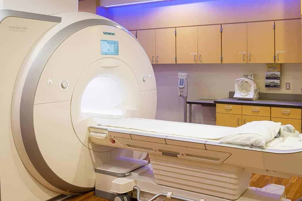
Magnetic Resonance Imaging (MRI) uses magnetic fields and radiofrequency waves to generate detailed pictures of internal structure. The MRI scan can show parts of the body not visible using a traditional X-ray and produces very clear three-dimensional images. Because of its safety and clarity, the MRI is a valuable tool that can aid in the diagnosis of a wide range of conditions. Bear in mind that you will be asked to remove all metal objects as a safety precaution. This includes: jewelry, watches, hearing aids and removable dental work.
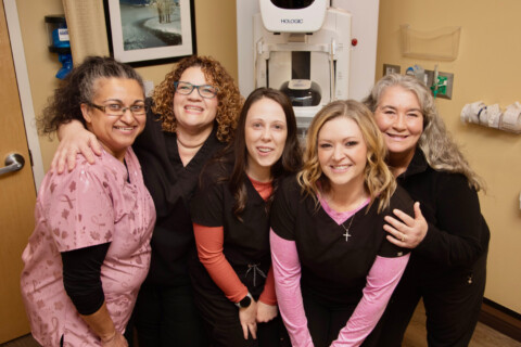
Mammography is the most common breast imaging tool that produces detailed images of the breast using specific X-ray technology. There are two types of mammography: screening and diagnostic. Screening mammograms are performed if you have no breast abnormalities or family history. Diagnostic mammograms are performed when you do have a symptom such as a lump or if an abnormality is found during the screening exam. Our goal is to detect cancers of the breast in the earliest stages and before a lump is ever felt.
Our new 3D mammogram is similar to the traditional mammogram; however, it reduces overlap in the breast images and uncovers distortion and speculated masses. This technology detects 20-65% more invasive breast cancers!
Nuclear Medicine is an imaging technique that uses small amounts of radioactive materials to diagnose and determine the severity of an illness. It can also be used to evaluate how well various parts of the body are functioning. Nuclear medicine tests are very sensitive and provide unique information that X-rays or ultrasounds cannot. Common nuclear medicine exams include bones, lungs, heart, thyroid and gallbladder. While the studies do use radiation, the dose is very low and is rapidly excreted from the body. Nuclear medicine studies require a physician referral.
Positron Emission Tomography (PET) is unique nuclear medicine technology that produces pictures of the body’s biological functions. PET studies have the ability to detect metabolic changes caused by diseases, such as cancer. PET scans can be performed on the entire body and gives your physician a wide range of information, enabling the best possible treatment plan. The PET scan requires a physician referral and has strict preparation guidelines.
Ultrasound scans create real-time images of the inside of the body using sound waves that are converted into electrical impulses. Ultrasounds do not use radiation and are generally painless and non-invasive. Ultrasound imaging can be used for screening, diagnosis or to assist with a treatment.

One of the goals I have as Chief Medical Officer at Island Health is to positively impact our community’s health…
Early detection saves lives! Island Health is proud to offer many preventive cancer screenings to help keep our community healthy.*…

“I can't say enough good things about the mammogram tech and radiologist. It was a painless process and they put me completely at ease during it all. I wonder sometimes if it is too dramatic to say my mammogram saved my life. But it might have!”

“Don’t ignore yourself. You know what feels wrong.”